Home
/ Back Bones Diagram - Upper Back Pain Anatomy Of The Back The Pain Center Pain Management Care / There are three parts to the trapezius.
Back Bones Diagram - Upper Back Pain Anatomy Of The Back The Pain Center Pain Management Care / There are three parts to the trapezius.
Back Bones Diagram - Upper Back Pain Anatomy Of The Back The Pain Center Pain Management Care / There are three parts to the trapezius.. Cervical bones diagram 12 photos of the cervical bones diagram cervical bones diagram, cervix anatomy. Bones of the back anatomy tutorials. The atlas is a ring of bone made up of two lateral masses joined at. It also covers some common conditions and injuries that can affect the back. The lumbar spine makes up the the lower end of the spinal column.
The anatomy of the lumbar spine is quite complex. The atlas is the topmost vertebra, and along with c2, forms the joint connecting the skull and spine. Human spine diagram reading industrial wiring diagrams. The spine anatomy is a complex structure. In the back and elsewhere in the body, tendons attach muscles to bones.
Back Muscles Anatomy Function Treatment from www.verywellhealth.com The spine anatomy is a complex structure. The bones of the pelvis and lower back work together to support the body's weight, anchor the abdominal and hip muscles, and protect the delicate vital organs of the vertebral and abdominopelvic cavities. Skeleton bone back human body human anchor chart health science diagram. Muscle or tendon injuries can occur anywhere in the body. Back to short bone diagram. Individual anatomical structures include 2: Human back bones diagram poster 28 inch x 24 inch 16 inch x 13 inch. Draw vertical arrows connecting the categories to the main backbone arrow.
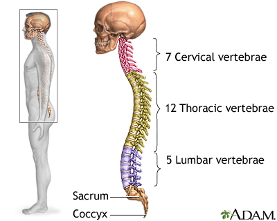
It consists of 5 lumbar vertebra that are numbered 1 through 5 from top to bottom i.e.
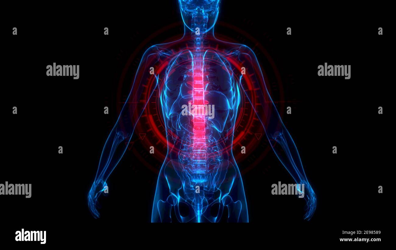
We hope this picture anatomy of back muscles diagram can help you study and research. Human back bones diagram poster 28 inch x 24 inch 16 inch x 13 inch. The bones of the pelvis and lower back work together to support the body's weight, anchor the abdominal and hip muscles, and protect the delicate vital organs of the vertebral and abdominopelvic cavities. Seven cervical vertebrae in the neck, twelve thoracic vertebrae in the torso and five lumbar vertebrae in the lower back. Spinal anatomy and back pain. See lumbar spine anatomy diagram stock video clips. Download 2,401 bones diagram stock illustrations, vectors & clipart for free or amazingly low rates! Your lower back contains 5 vertebral bones stacked above each other with intervertebral discs in between. It contains the osteology, arthrology and myology of the spine and back. Bones, discs, and joints in your lower back. The atlas is a ring of bone made up of two lateral masses joined at. Its appearance is different from the other spinal vertebrae. Bones of the back anatomy tutorials.
Related posts of human back bones diagram pelvic bone labeled. Its appearance is different from the other spinal vertebrae. The anatomy of the lumbar spine is quite complex. If the cause is large or complex, it is best to. Seven cervical vertebrae in the neck, twelve thoracic vertebrae in the torso and five lumbar vertebrae in the lower back.
3d Spine High Resolution Stock Photography And Images Alamy from c8.alamy.com Anatomynote.com found anatomy of back muscles diagram from plenty of anatomical pictures on the internet. The occiput (co), also known as the occipital bone, is a flat bone that forms the back of the head. The spine supports your body and helps you walk, twist and move. This vertebra supports the skull. The vertebrae, which stack like spools of thread, support the back and protect the spinal cord. Draw vertical arrows connecting the categories to the main backbone arrow. Its appearance is different from the other spinal vertebrae. We think this is the most useful anatomy picture that you need.
12 photos of the human back bone chart.
Human back bones diagram poster 28 inch x 24 inch 16 inch x 13 inch. Hip bones diagram of back and hip bones 9 out of 10 based on 30 ratings. They help support particular bones and make them move. The vertebrae, which stack like spools of thread, support the back and protect the spinal cord. Fishbone diagrams, aka ishikawa diagrams are used across various industries to analyze causes and their effect. Human backbone diagram, bone, human backbone diagram. Spinal vertebrae bone spine vertebra toracica spinal cord spine structure back diagram spine sections spinal cord vertebrae spinal structure health diagram. Your lower back contains 5 vertebral bones stacked above each other with intervertebral discs in between. This article looks at the anatomy of the back, including bones, muscles, and nerves. 12 photos of the human back bone chart. The occiput (co), also known as the occipital bone, is a flat bone that forms the back of the head. The lower part of the trapezius ascends and depresses the scapula, while the transverse or middle region of the trapezius is what retracts the. The lumbar spine is the lower back that begins below the last thoracic vertebra (t12) and ends at the top of the sacral spine, or sacrum (s1).
Draw vertical arrows connecting the categories to the main backbone arrow. Bones of the pelvis and lower back. Stress fracture treatment symptoms u0026 causes. We hope this picture anatomy of back muscles diagram can help you study and research. Topographic anatomy of the back the lecturio medical online library.
Skeletal Spine Medlineplus Medical Encyclopedia Image from medlineplus.gov The lower part of the trapezius ascends and depresses the scapula, while the transverse or middle region of the trapezius is what retracts the. The spine diagram the spine diagram shown below, consists of many bones or vertebrae,soft discs,the spinal cord, and spinal nerves. The atlas is a ring of bone made up of two lateral masses joined at. The l5 vertebra is connected to the top of. Each typical vertebra consists of a body, an arch and three processes that stem from. In the back and elsewhere in the body, tendons attach muscles to bones. Bones front and back diagram quizlet : Over 3000+ pages with full illustrations and diagrams.
The vertebral column of the lower back includes the five lumbar vertebrae, the sacrum, and the coccyx.
These bones are connected at the back with specialized joints. We hope this picture anatomy of back muscles diagram can help you study and research. Created and produced by qa international. The occiput (co), also known as the occipital bone, is a flat bone that forms the back of the head. 12 photos of the human back bone chart. For more anatomy content please follow us and visit our website: Bones of the back anatomy tutorials. The red lines point individual bones and the names are writen in singular, the blue lines conect to group of bones and are in plural form. Thorax,lungs,heart anatomy and physiology diagrams free download. Pelvic bone labeled 12 photos of the pelvic bone labeled pelvic bone labeled, pelvic bone labeling quiz, pelvic bone with labeling, pelvic girdle bone labeling quiz, pubic bone labeled, bone, pelvic bone labeled, pelvic bone labeling quiz, pelvic bone with labeling, pelvic girdle bone labeling quiz, pubic bone labeled At the same time the bones grow larger by growing back into the growth plates. The bones of the appendicular skeleton provide support and flexibility at the joints and anchor the muscles that move the limbs. But on the inside there are many similarities among human, bird, and bat forearms.
The vast difference in height and limb length between birth and adulthood are mainly the result of endochondral ossification in the back bones. But on the inside there are many similarities among human, bird, and bat forearms.
:max_bytes(150000):strip_icc()/GettyImages-56372373-1bb8a07f23d84f439c3e80ecf3c472d2.jpg)
:max_bytes(150000):strip_icc()/GettyImages-56372373-1bb8a07f23d84f439c3e80ecf3c472d2.jpg&description=Back Bones Diagram - Upper Back Pain Anatomy Of The Back The Pain Center Pain Management Care / There are three parts to the trapezius.)